
Software
YT-SOFTWARE ecosYsTem
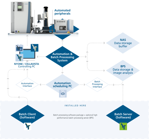
SYNENTEC provides in the field of automated imaging systems the latest standard in image acquisition, device controlling and image- and data analysis software. We offer a solution for label-free, high-throughput image analysis using artificial intelligence (AI) models. And the available data batch-processing solutions provide highest claims in plate throughput - 21 CFR part 11 / Annex 11 compliant.
Discover all the advantages
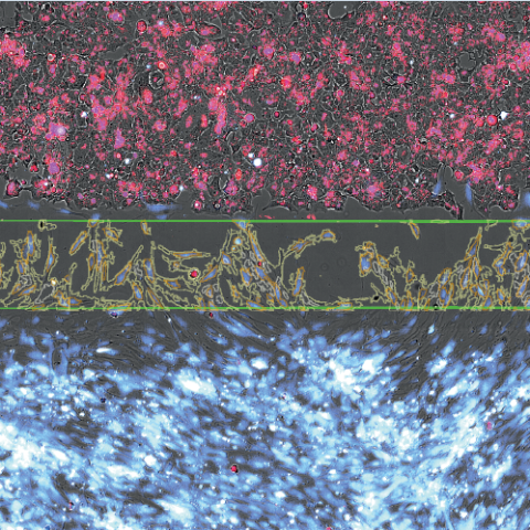
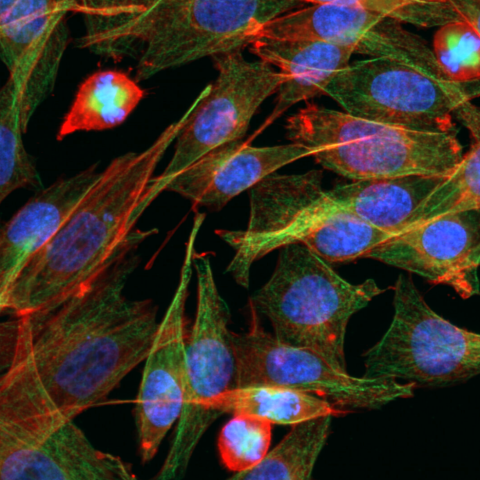
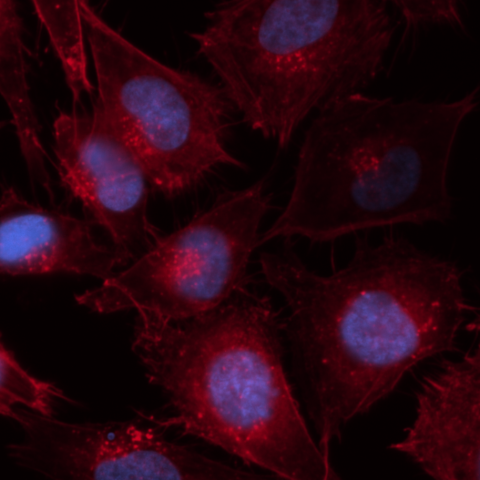
The Future is bright and fluorescent
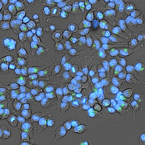
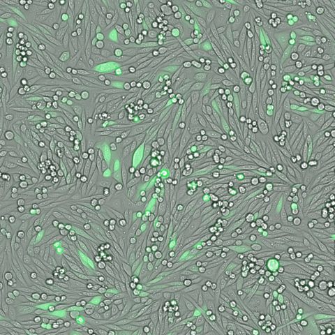
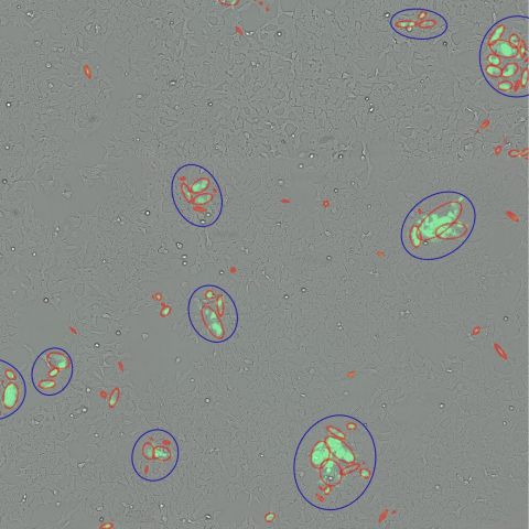
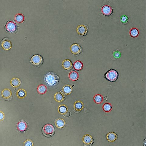
- AI-based staining free viability
- Single Cell Cloning
- Trypan Blue Testing
- Viral Plaque Assay (non invasive)
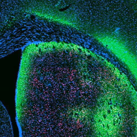
- iPS Cells on Feederlayer
- Cornea Cell Count
- Adherent Cell Proliferation
- SiRNA Detection
- Transfection Efficiency
- Wound Healing & Scratch Assay
- FASCC (Cloning)
- Transfection & CRISPR/Cas9
- Rare Cell Detection
- Spheroid Imaging
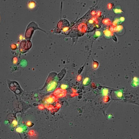
- AnnexinV Apoptosis
- Viral Infectivity
- Seeding Method Validation (e.g. FACS)
- Caspase Assay
- PAIA igG & FC Quantitation
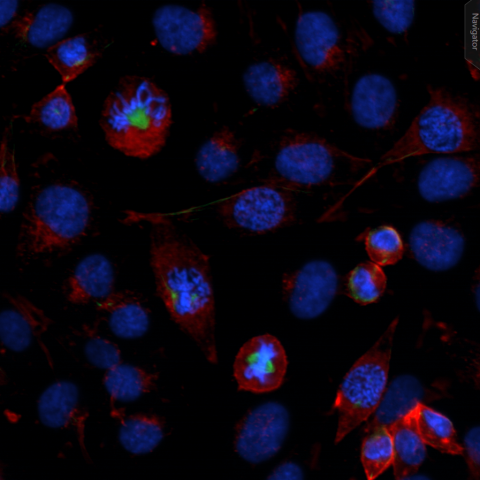
- Cell Cycle & Mitosis
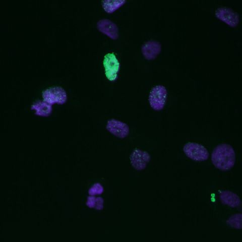
- yH2AX DNA Damage
- ICC & Multiplexing
- JC-1 Apoptosis
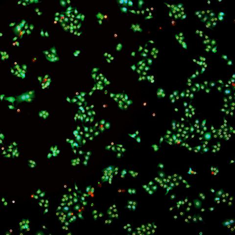
- LIVE/DEAD Toxicity Study
- HCS Assays
- Oxidative Stress / ROS
The Future is bright and fluorescent
DO YOU WANT TO KNOW MORE?
We know that time is an increasingly scarce resource, even in the lab. That's why we've thought your problem through and have everything ready for a complete one-handed solution.