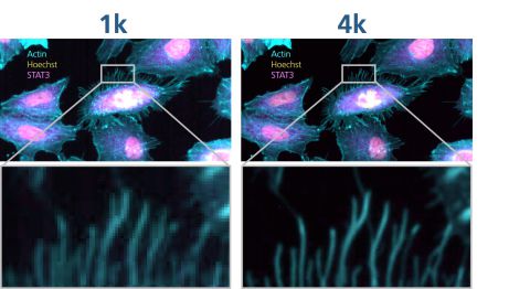
High content screening using 4K resolution
COMPARE TECHNICAL SPECIFICATIONS
|
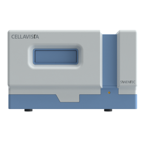
CELLAVISTA® 4K HighEnd1 |
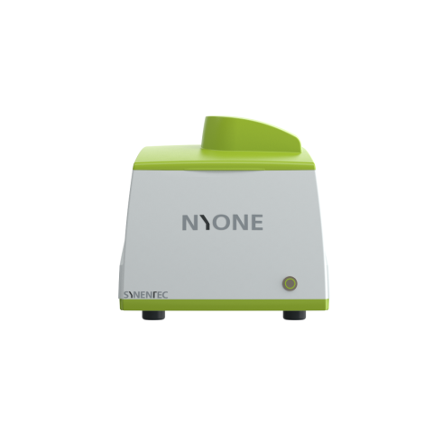
NYONE® 4K HighEnd1 |
CELLAVISTA® 4 Scientific |
NYONE® Scientific |
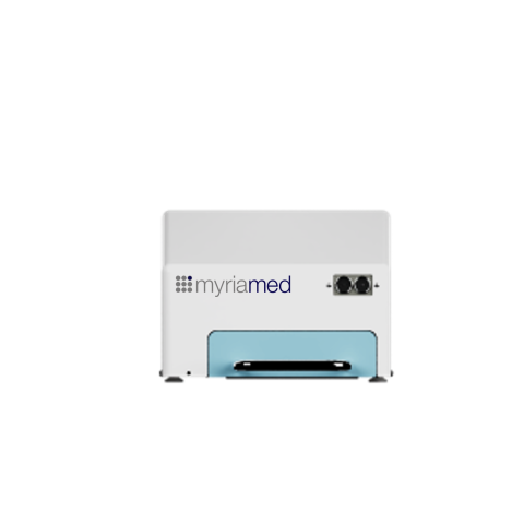
myrImager |
|
|---|---|---|---|---|---|
|
Throughput |
250² plates / day |
100² plates / day |
250² plates / day |
150² plates / day |
48 samples simultaneously (during desired observation period) |
|
Objective capacity |
4 |
3 |
4 |
3 |
1 |
|
Selectable Resolutions1 |
3.3 µm @ 2x optional |
1.3 µm @ 4x |
6.5 µm @ 2x optional |
6.5 µm @ 2x optional |
0.086x magnification |
|
Automated whole well/whole plate imaging |
✓ |
✓ |
✓ |
✓ |
whole plate one shot |
|
Sample types |
SBS plate format (1536, 384, 96 wells and less), culture dishes and microscope slides |
SBS plate format (1536, 384, 96 wells and less), culture dishes and microscope slides |
SBS plate format (1536, 384, 96 wells and less), culture dishes and microscope slides |
SBS plate format (1536, 384, 96 wells and less), culture dishes and microscope slides |
myrPlates |
|
Camera |
8 bit progressive scan |
8 bit progressive scan |
16 bit sCMOS |
16 bit sCMOS |
8 bit progressive scan |
|
Pixel density |
5440x 5440 |
4496 x 4496 |
2048 x 2048 |
2048 x 2048 |
3088 x 2064 |
|
Quantum efficiency |
~57 % |
~66 % |
> 80 % |
~80 % |
80 % |
|
Light source |
High performance long-life LED |
High performance long-life LED |
High performance long-life LED |
High performance long-life LED |
Long-life LED |
|
Illumination / Fluorescence1 |
Brightfield and 6 fluorescence excitation/emission channels |
Brightfield and 4 fluorescence excitation sources, up to 5 fluorescence emission filters |
Brightfield and 6 fluorescence excitation/emission channels |
Brightfield and 4 fluorescence excitation sources, up to 5 fluorescence emission filters |
Long-life LED |
|
Temp.-& CO2 control |
Possible3 |
Possible3 |
Possible3 |
Possible3 |
24/7 incubator integration |
|
Image acquisition- & device controlling software4 |
YT-SOFTWARE®4 |
YT-SOFTWARE®4 |
YT-SOFTWARE®4 |
YT-SOFTWARE®4 |
YT-myrImage |
|
Automation-ready set up |
✓ |
✓ |
✓ |
× |
N.A. |
|
Software-interface for automation |
✓ |
✓ |
✓ |
✓ |
N.A. |
|
Batch processing interface |
✓ |
✓ |
✓ |
✓ |
N.A. |
|
External barcode-reader |
Optional |
Optional |
Optional |
Optional |
Optional |
1 Different system configurations like e.g. basic systems with brightfield only or customized filter and objective lenses set-ups are available.
2 Throughput per day depends on experiment configurations (e.g. exposure time, plate type, focus options, # of imaging channels…).
3 CELLAVISTA® & NYONE® can be combined with an external incubation and plate handling option (SYBOT X-1000 or other automation systems), whereby the plates are only outside the incubator during the measuring time.
4 YT-SOFTWARE® includes the same image analysis tools for both CELLAVISTA® and NYONE®. Differences in usable applications arise only due to different hardware configurations (e.g. different illuminations or camera chips).
5 Hardware automation-package available.
DO YOU WANT TO KNOW MORE?
We know that time is an increasingly scarce resource, even in the lab. That's why we've thought your problem through and have everything ready for a complete one-handed solution.


