
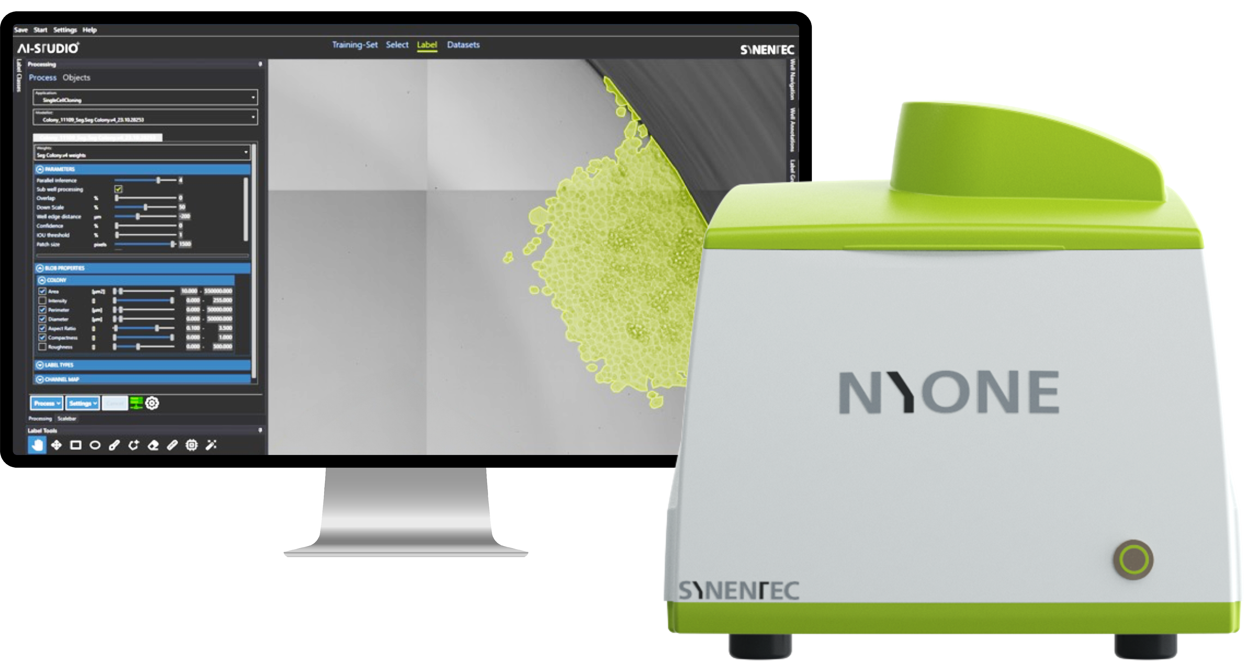
NYONE
Compact Image Cytometry
This fully automated cell imager and image analysis device is characterized by its mechanical robustness, small footprint, optical quality and functionality balanced features in a way that you will soon recognize that NYONE® is a valuable member of your team - ideal partner for medium-throughput laboratories with limited bench space!
Discover all the advantages
COMPARE TECHNICAL SPECIFICATIONS
|
NYONE® 4K HighEnd1 |
NYONE® Scientific |
|
|---|---|---|
|
Throughput |
100² plates / day |
150² plates / day |
|
Objective capacity |
3 |
3 |
|
Selectable Resolutions1 |
1.3 µm @ 4x |
6.5 µm @ 2x optional |
|
Automated whole well/whole plate imaging |
✓ |
✓ |
|
Sample types |
SBS plate format (1536, 384, 96 wells and less), culture dishes and microscope slides |
SBS plate format (1536, 384, 96 wells and less), culture dishes and microscope slides |
|
Camera |
8 bit progressive scan |
16 bit sCMOS |
|
Pixel density |
4496 x 4496 |
2048 x 2048 |
|
Quantum efficiency |
~66 % |
~80 % |
|
Light source |
High performance long-life LED |
High performance long-life LED |
|
Illumination / Fluorescence1 |
Brightfield and 4 fluorescence excitation sources, up to 5 fluorescence emission filters |
Brightfield and 4 fluorescence excitation sources, up to 5 fluorescence emission filters |
|
Temp.-control |
× |
× |
|
Image acquisition- & device controlling software4 |
YT-SOFTWARE®4 |
YT-SOFTWARE®4 |
|
Automation-ready set up |
✓ |
× |
|
Software-interface for automation |
✓ |
✓ |
|
Batch processing interface |
✓ |
✓ |
|
External barcode-reader |
Optional |
Optional |
1 Different system configurations like e.g. basic systems with brightfield only or customized filter and objective lenses set-ups are available.
2 Throughput per day depends on experiment configurations (e.g. exposure time, plate type, focus options, # of imaging channels…).
3 CELLAVISTA® can be equipped with the ibidi® heating and incubation system (other systems not tested). Due to pH-shift and consequent cell stress a heating option only is not recommended.
4 YT-SOFTWARE® includes the same image analysis tools for both CELLAVISTA® and NYONE®. Differences in usable applications arise only due to different hardware configurations (e.g. different illuminations or camera chips).
5 Hardware automation-package available.


Advanced Technological Features
- Agitation-free imaging of adherent and suspension cells through harmonic motion
- Reliably reproducible measurements through high-precision scan stage control
- Ultra-fast filter wheel – 40 ms position changing time (e.g. well suited for FRET assays)
- Excellent well edge illumination of microplates
- Perfect, seemless image stitching
- Ultra-fast electronically switched excitation sources - less than 5 ms switching time
- Always in focus due to specially designed laser autofocus mechanisms
- Leverages the flexibility of automated image analysis to maximize the output of your cellular assays
To get an overview compare all high throughput microscopes >>>
DO YOU WANT TO KNOW MORE?
We know that time is an increasingly scarce resource, even in the lab. That's why we've thought your problem through and have everything ready for a complete one-handed solution.